Beyond Cirrhosis: Refining HCC Risk Stratification in MASLD
18 Nov 2026
Share
The increasing contribution of metabolic dysfunction-associated steatotic liver disease (MASLD) to the hepatocellular carcinoma (HCC) burden highlights the challenge of identifying at-risk patients who warrant surveillance.1,2 Current HCC surveillance strategies largely focus on patients with cirrhosis, yet HCC can develop in patients with MASLD before cirrhosis is clinically recognized.3,4 However, extending surveillance to the entire MASLD population would place substantial demands on healthcare resources.3 In an interview with Omnihealth Practice, Professor Chan, Wah Kheong, and Professor Yip, Cheuk-Fung Terry, discussed how sequential assessment using the non-invasive Fibrosis-4 index (FIB-4), followed by liver stiffness measurement (LSM), could enable more targeted HCC risk stratification in MASLD. This approach may help identify patients who warrant further evaluation for surveillance while avoiding unnecessary assessment of those at lower risk.
A changing HCC landscape calls for better risk stratification
MASLD affects more than 30% of the general population and is an increasingly important cause of HCC.5 Prof. Chan described a shifting HCC landscape in Malaysia, where recent data from his center suggest that MASLD has overtaken chronic hepatitis B as the leading etiology among newly diagnosed HCC cases (48% vs. 32%, respectively).6 In Hong Kong, Prof. Yip noted that viral hepatitis remains predominant, accounting for approximately 80% of HCC cases, with metabolic risk factors and MASLD increasingly contributing to the burden.7
The key challenge is identifying patients whose HCC risk is sufficiently high to warrant surveillance.1 Prof. Chan emphasized: “Early detection of HCC depends on appropriate surveillance of the at-risk population.” Current guidelines generally recommend surveillance for patients with MASLD-related cirrhosis. An annual HCC incidence of ≥1% has been proposed as a threshold for cost-effective surveillance in patients with cirrhosis, regardless of etiology.3,8,9 However, a cirrhosis-based approach may leave some patients with MASLD outside established surveillance pathways, as up to 30%-50% of MASLD-related HCC cases occur without established cirrhosis, and underlying cirrhosis may remain unrecognized.8,10
Further complicating risk stratification, HCC incidence is lower in MASLD than in other chronic liver diseases, including among patients with cirrhosis.2,8 Prof. Chan also noted that hepatic steatosis may obscure typical sonographic features of cirrhosis, delaying recognition of advanced liver disease until patients present with decompensation.8,10 Together, these considerations underscore the limitations of relying on a binary cirrhosis vs. non-cirrhosis classification and support more granular assessment of fibrosis severity and individual HCC risk.1
Sequential FIB-4 and LSM assessment refines HCC risk stratification
The value of a more granular approach to HCC risk stratification has been demonstrated in a multicenter study of 12,950 patients with MASLD followed for a median of 47.7 months.8 HCC incidence increased across higher FIB-4 and LSM categories (figure 1).8 FIB-4 showed a clear gradient in HCC risk, with annual HCC incidence increasing from 0.07% among patients below the age-specific low-risk threshold (FIB-4 <1.3 for those aged <65 years and <2.0 for those aged ≥65 years) to 1.18% among those with FIB-4 ≥3.25 (figure 1A).8 LSM showed a similar gradient, with annual HCC incidence reaching ≥1% among patients with LSM ≥20kPa (figure 1B).8
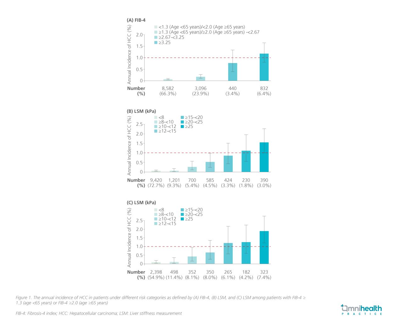
These findings support a sequential approach, using FIB-4 for initial assessment and LSM to further stratify patients above the age-specific low-risk FIB-4 threshold.8 As standalone measures, FIB-4 ≥3.25 and LSM ≥20kPa were independently associated with an annual HCC incidence exceeding the HCC surveillance threshold.8 However, among patients with FIB-4 above the age-specific low-risk threshold, an LSM threshold of ≥15kPa identified a group with an annual HCC incidence ≥1% (figure 1C).8 This suggests that sequential assessment may identify additional at-risk patients with LSM 15kPa to <20kPa who might not be identified as high risk using LSM alone.8
Prof. Chan explained that “sequential use of FIB-4 and LSM can improve risk stratification while conserving resources by avoiding the need to perform LSM in all patients.” By enriching the LSM population through initial FIB-4 assessment, this approach may improve the yield of subsequent LSM while enabling more targeted use of healthcare resources. Prof. Yip similarly highlighted that “Stepwise risk assessment provides a practical balance between clinical utility and accuracy.” FIB-4 also facilitates initial risk assessment beyond hepatology settings, as it relies on routinely available parameters: age, aspartate aminotransferase (AST), alanine aminotransferase (ALT, and platelet count.
Translating risk stratifications into clinical practice
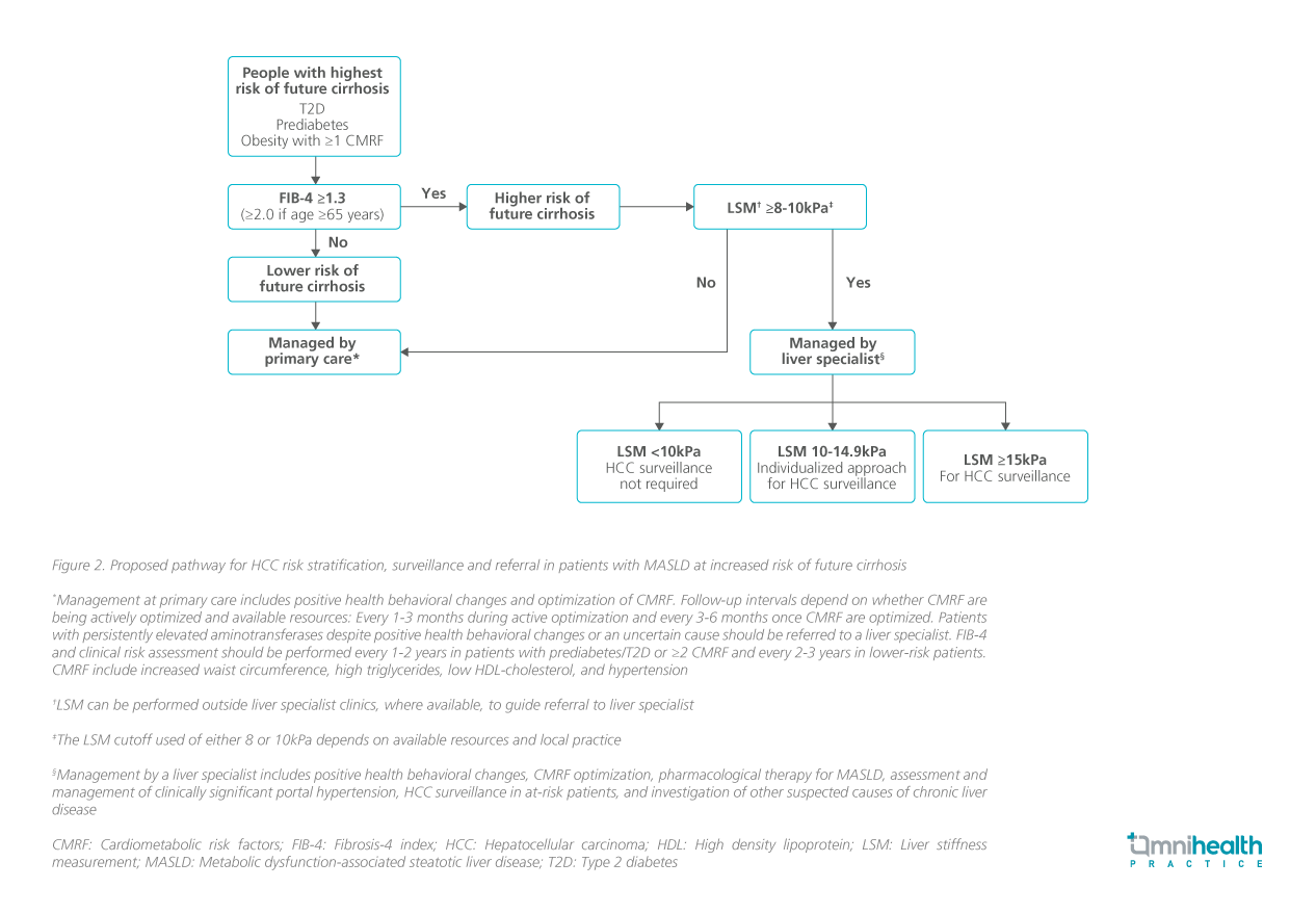
Although the proposed thresholds provide a practical framework for HCC risk stratification, Prof. Chan emphasized that non-invasive measurements should be interpreted within the broader clinical context rather than as absolute determinants of surveillance eligibility. Among patients with an elevated FIB-4, an LSM <10kPa may indicate a lower HCC risk, whereas an LSM of 10-14.9kPa represents an intermediate-risk range requiring individualized surveillance decisions based on additional clinical features, including platelet count and other evidence of advanced liver disease.9 An LSM ≥15kPa identifies a higher-risk group in whom HCC surveillance should be considered in the context of overall clinical picture.9
These risk thresholds help inform the respective roles of primary and specialist care. Prof. Chan noted that “risk stratification can help direct patients with more advanced liver disease to specialist care, while allowing those with less severe disease to remain under primary care or endocrinology.” Primary care can initiate FIB-4 assessment, followed by LSM where available to refine risk stratification before specialist referral.9,11 This approach may focus specialist resources on patients with greater evidence of advanced liver disease and guide surveillance and referral decisions through a sequential FIB-4 and LSM pathway (figure 2).9,11
Longitudinal reassessment of fibrosis and HCC risk
HCC risk assessment should be viewed as a dynamic longitudinal process, as metabolic and fibrosis risk may change over time. Prof. Yip noted that most patients with MASLD would not require HCC surveillance at baseline, and those with a low FIB-4 could generally remain in non-hepatology care. He emphasized that “periodic FIB-4 reassessment at approximately 1-3-year intervals is important for identifying changes in risk that may warrant further assessment with LSM or specialist evaluation, as well as reconsideration of HCC surveillance eligibility.”8,11
Notably, changes in fibrosis measures should not automatically be interpreted as changes in HCC risk or as a basis for de-escalating surveillance. As the study assessed FIB-4 and LSM only at baseline, it does not establish whether longitudinal changes reliably reflect HCC risk or should guide surveillance decisions.8 Prof. Chan explained that newer pharmacological therapies for MASLD can produce substantial weight loss and reductions in LSM, but these improvement may not necessarily indicate a sufficient reduction in HCC risk to justify discontinuing surveillance, particularly in patients with underlying advanced liver disease.
Bridging the gap between risk identification and clinical action
Identifying high-risk patients is only the first step; effective surveillance requires translating risk assessment into clinical action.9 Prof. Chan emphasized the value of early HCC detection, noting that it increases the opportunity for curative treatment and is associated with improved survival and cost-effectiveness.8,9
However, Prof. Chan highlighted the implementation gap in clinical practice, noting that, in one diabetes clinic, approximately 28% of patients had an elevated FIB-4, yet only around 12% were referred for further evaluation. In a separate multicenter randomized study conducted in Malaysia and Hong Kong, automated fibrosis-score calculation with electronic reminders increased appropriate care among patients with increased fibrosis scores from 3.1% to 33.3%, although more than half still received no further assessment or referral.12
Clear referral criteria and defined roles across primary care, endocrinology and hepatology are essential for a coordinated pathway.9 Addressing patient-, clinician- and system-level barriers to surveillance, as outlined in table 1, is similarly important to translate risk-based HCC surveillance into routine MASLD care.9
| Patient-level barriers | Clinician-level barriers | System-level barriers |
| Low awareness of liver disease and HCC risk | Time and workload constraints | Limited surveillance infrastructure |
| Socioeconomic and logistical barriers to healthcare | Underrecognition of surveillance eligibility in primary care | Maldistribution of specialists and workforce |
| Stigma and discrimination | Lack of integrated tracking and recall systems |
Table 1. Patient, clinician, and system-level barriers to effective HCC surveillance
HCC: Hepatocellular carcinoma
Further evidence is needed to define the surveillance pathway
Further evidence is needed before risk-based surveillance can be more broadly implemented. Prof. Yip emphasized the need for prospective validation, longer-term follow-up and health economic analyses to confirm whether the approximately 1% annual HCC incidence observed in higher-risk patients is reproducible across clinical settings and whether the two-step strategy improves outcomes in routine practice.
The optimal surveillance modality also warrants further consideration. Prof. Yip highlighted the limited sensitivity of conventional ultrasound and alpha-fetoprotein (AFP) for HCC detection.1,3 Magnetic resonance imaging may have a role in selected higher-risk populations, although its greater resource requirements remain a consideration.1,3 Biomarker-based approaches such as GALAD (gender, age, alpha-fetoprotein L3 [LAF], alpha-fetoprotein [AFP], des-gamma-carboxy prothrombin [DCP]) also warrant further investigation.2 As risk-based strategies may extend surveillance to selected patients without established cirrhosis, the optimal surveillance approach must balance diagnostic performance, clinical feasibility and resource requirements.1,8
Conclusion
HCC risk in MASLD extends beyond a binary cirrhosis-based classification, supporting a more granular approach to fibrosis and HCC risk stratification.8 Prof. Chan emphasized the value of assessing fibrosis severity to identify patients who may warrant closer evaluation or surveillance, while Prof. Yip highlighted the potential of non-invasive risk stratification to move beyond a cirrhosis-only surveillance paradigm. Sequential FIB-4 and LSM assessment may help identify patients for further evaluation, surveillance considerations and specialist referral, while enabling more targeted use of healthcare resources.8 Implementation will require longitudinal reassessment, accessible LSM, clear referral and surveillance pathways, alongside further prospective validation and health economic evidence.8,9
References
- European Association for the Study of the Liver (EASL), European Association for the Study of Diabetes (EASD), European Association for the Study of Obesity (EASO). EASL-EASD-EASO Clinical Practice Guidelines on the Management of Metabolic Dysfunction-Associated Steatotic Liver Disease (MASLD). Obes Facts. 2024;17:374-443.
- Rich NE, et al. AGA Clinical Practice Update on Risk Stratification and Emerging Surveillance Strategies for Hepatocellular Carcinoma: Expert Review. Gastroenterology. 2026;170(7):1606-1615.
- Singal AG, et al. AASLD Practice Guidance on prevention, diagnosis, and treatment of hepatocellular carcinoma. Hepatology. 2023;78(6):1922-1965.
- Worthington J, et al. Introducing risk stratification reduces patient burden and improves cost-effectiveness of hepatocellular carcinoma surveillance for people with metabolic dysfunction-associated steatotic liver disease: a modeling study. Value Health. 2026. doi:10.1016/j.jval.2026.06.018.
- Younossi ZM, et al. Epidemiology of metabolic dysfunction-associated steatotic liver disease. Clin Mol Hepatol. 2025;31(Suppl):S32-S50.
- Teoh E-S, et al. Hepatocellular carcinoma in Malaysia: Shifting trend in aetiology and the impact of surveillance on clinical outcomes. Presented at the Malaysian Society of Gastroenterology and Hepatology (MSGH) GUT Annual Scientific Meeting 2026; August 28-30, 2026.
- Zhu RX, et al. Epidemiology of hepatocellular carcinoma in the Asia-Pacific region. Gut Liver. 2016;10(3):332-339.
- Lai JC-T, et al. Non-invasive risk-based surveillance of hepatocellular carcinoma in patients with metabolic dysfunction-associated steatotic liver disease. Gut. 2025;74(12):2050-2057.
- Lau SY, et al. The Malaysian Society of Gastroenterology and Hepatology consensus statements on prevention and early detection of hepatocellular carcinoma in Malaysia. J Gastroenterol Hepatol. 2026;41(9):2639-2655.
- Bertot LC, et al. Nonalcoholic fatty liver disease-related cirrhosis is commonly unrecognized and associated with hepatocellular carcinoma. Hepatol Commun. 2017;1(1):53-60.
- Kanwal F, et al. Clinical care pathway for the risk stratification and management of patients with metabolic dysfunction-associated steatotic liver disease. Gastroenterology. 2026;171(1):164-183.
- Zhang X, et al. Clinical care pathway to detect advanced liver disease in patients with type 2 diabetes through automated fibrosis score calculation and electronic reminder messages: A randomised controlled trial. Gut. 2023;72(12):2364-2371.